¶ Key Understandings:
- All living organisms are composed of cells, the basic unit of life. Various structures within the cell perform different functions.
- Cells are grouped into higher levels of organisation to allow for specialisation and division of labour.
- State that all organisms are composed of cells, which are the fundamental units of life.
- Differentiate cell, tissue, organ and organ system.
- Identify cell structures (including organelles) of typical plant and animal cells from diagrams, electron micrographs, and as seen under the light microscope.
- State the characteristics and functions of the components of protoplasm.
- State the characteristics and functions of the organelles and membrane structures found in the cell.
- Compare the structure of typical animal and plant cells.
- State the differences between prokaryotic and eukaryotic cells.
- state the benefits of division of labour in multicellular organisms
- Describe the relationship between cell function and cell structure of red blood cells in transport of oxygen, root hair cells in absorption, and epithelial cells of the small intestines in absorption.
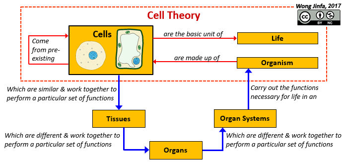
¶ Cell Theory (Schleiden, Schwann and Virchow, 1938)
- All living organisms are composed of one or more cells
- The cell is the most basic unit of life
- Cells display the characteristics of life
- Cells are the building blocks of life
- All cells come from pre-existing cells
¶ Differentiate cell, tissue, organ and organ system.
- A group of similar cells which work together to perform a particular set of function(s) are known as tissues
- Simple tissues are made up of only one type of cells
- Complex tissues are made up of several types of cells
- Different tissues working together to carry out a particular set of functions make up an organ
- Different organs working together to carry out a particular set of functions make up an organ system
¶ Identify cell structures (including organelles) of typical plant and animal cells from diagrams, electron micrographs, and as seen under the light microscope.
- Cells are too small to be seen by the naked eye and require microscopes to be seen
- Robert Hooke was the first to observe cells of thin slices of bottle cork under the microscope in 1665
- Cameras can be fitted to the microscope to take pictures called micrographs
- Cells can be viewed from different perspectives:
- Longitudinal Section: Cutting along the long axis of the cell
- Transverse Section: Cutting at right angles to the longitudinal plane
- Light Microscope
- Model found in most schools
- Use compound lenses to magnify objects
- The lenses bend or refract light to make the object beneath them appear closer
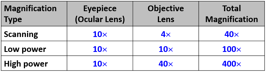
- The total magnification is the the product of the eyepiece magnification and the objective lens magnification
- Stereo Microscope
- This microscope allows for binocular (two eyes) viewing of larger specimens
- Scanning Electron Microscope (SEM)
- Allow scientists to view specimens too small to be seen with a light microscope
- SEMs do not use light waves; they use electrons (negatively charged electrical particles) to magnify objects up to two million times
- Transmission Electron Microscope (TEM)
- Also uses electrons, but instead of scanning the surface (as with SEM's) electrons are passed through very thin specimens
- Rotate the nosepiece of the microscope until the scanning objective lens (4x) clicks into position
- Look through the eyepiece and use the coarse adjustment knob to look for the specimen
- Once you spotted the specimen, switch to the lower power objective lens (10x) and use the coarse adjustment knob to refocus
- Switch to high power lens and use the fine adjustment knob to focus
- If the specimen is too light or too dark, try adjusting the diaphragm
- If you see a line in your viewing field, try twisting the eyepiece and the line should move (That is a pointer to point out features of the specimen)
- Please click on this link to practise focusing specimens using a virtual microscope
- Cut a thin piece of your specimen
- Place it on a microscope slide
- Place one drop of water directly over the specimen and spread the specimen so that no parts fold or overlap
- Add a drop of dye to stain the specimen if required
- Place the cover slip at a 45 degree angle with one edge touching the water drop and gently let it go with a mounting needle
- Blot the sides of the slide dry with a paper towel
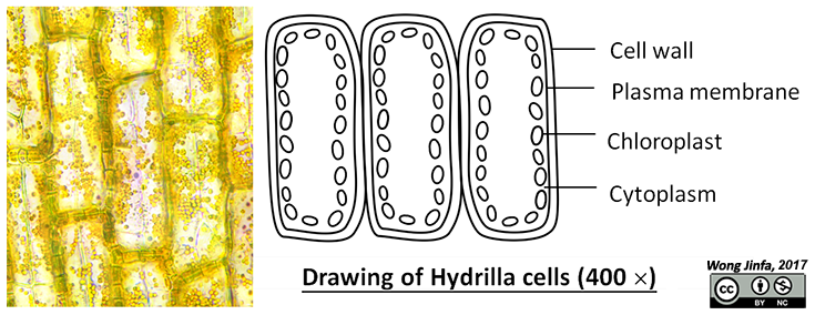
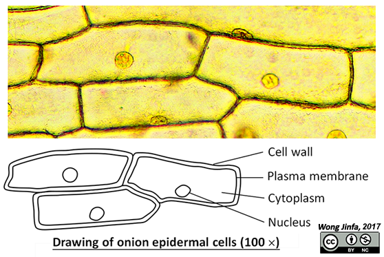
- Draw an outline (2D drawing) in pencil
- Use only discrete single lines; do not sketch or shade
- The size of the structures should be proportional
- At least 50% of the drawing space
- Draw label lines with a ruler, without an arrowhead
- Label lines should spread out radially from the drawing with the labels written outside the drawing
- Title should be underlined and include the magnification of the drawing
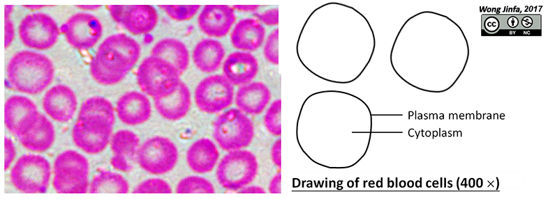
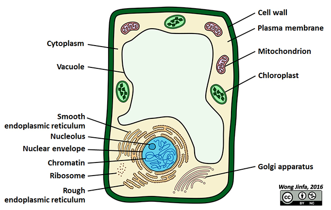
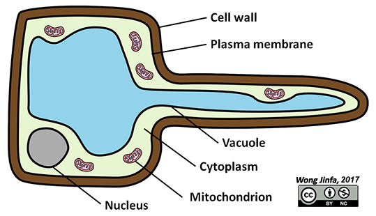
- The cell surface membrane or plasma membrane marks the boundary of a cell
- The fluid component bound by the plasma membrane (which is not a structure) is the cytoplasm
- The intracellular structures that you can observe under the light microscope include the nucleus and vacuole and chloroplasts (in plants)
- In plants, there is an extracellular structure that is found outside the plasma membrane known as the cell wall
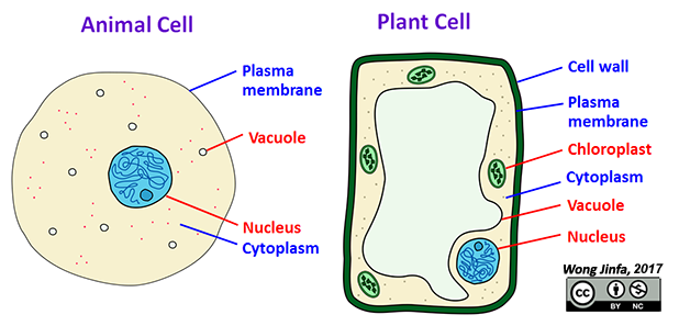
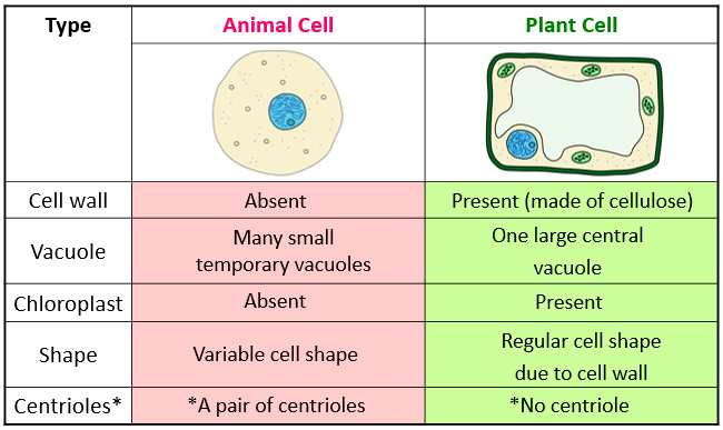
- Animal Cell:
- Plant Cell:
- Click here to see more electron micrographs by H. Jastrow
- Click on this link for a game created by Sheppard Software
¶ State the characteristics and functions of the components of protoplasm.
- The living matter that make up a cell is called protoplasm
- Protoplasm of cell is made up of three parts:
- Cell surface membrane / Plasma membrane
- Cytoplasm
- Nucleus
- The membrane that surrounds the entire cell
- The plasma membrane is made up of a phosopholipid bilayer with different types of proteins embedded in it
- Selectively/partially permeable
- Allows certain substances to pass through but not others (NOT semi-permeable)
- Functions:
- Controls the movement of dissolved substances in and out of the cell
- Separates the cellular contents from the external environment
- A gel-like aqueous fluid that fills the cell
- Contains dissolved molecules (e.g. proteins, sugars) and many specialised structures called organelles
- Cytosol refers to the fluid only (excluding the organelles)
- Function:
- Site for many chemical reactions and cellular activities
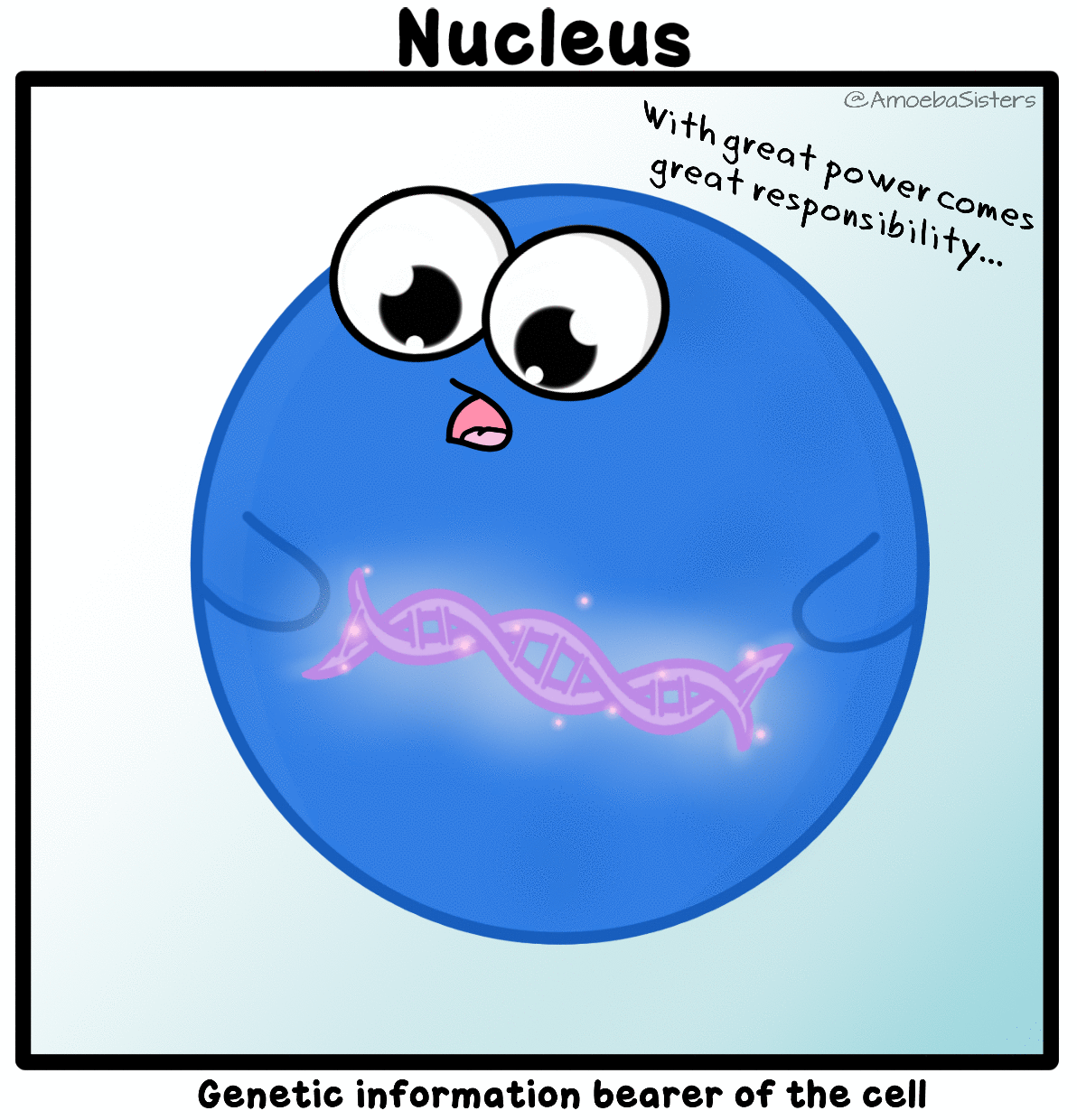
- Largest organelle in most cells
- Nuclear Envelope
- Nucleus is surrounded by a double membrane known as the nuclear envelope
- Outer membrane of the nuclear envelope is continuous with the endoplasmic reticulum
- Is perforated by nuclear pores, which allow exchange of substances between nucleus and the cytoplasm
- Nuceloplasm
- Gel-like matrix within the nucleus
- May contain one or more nucleoli and chromatin
- Nucleolus
- Contains large amount of DNA (deoxyribonulceic acids) & RNA (ribonucleic acids) which makes it stain deeply when a electron micrograph is taken
- Is the site of ribosome synthesis
- During nuclear division: nucleoli disappear as DNA disperse but they reassemble after nuclear division
- Chromatin
- Is made up of strands of deoxyribonucleic acid, DNA,wound around proteins
- Become highly coiled and condensed structures called chromosomes at the start of cell division
- (containing genetic/hereditary information)
- Functions:
- Contains the hereditary genetic materials of a cell in the form of DNA
- Is the control centre of all life processes of the cell (as DNA provides the instructions for protein synthesis)
- Involved in the production of ribosomes and ribonucleic acid
- Is an extracellular layer found in plant cells which is absent in animal cells
- The plant cell wall is made of cellulose (cf. chitin in fungi and peptidoglycan in bacteria)
- The cell wall is fully permeable
- Rigid (but flexible) shape acts as internal support for the plant, giving it a regular shape and protecting the cell from injuries
- Plasmodesmata are tiny channels that cut across plant cell walls, to allow transport and communication between cells
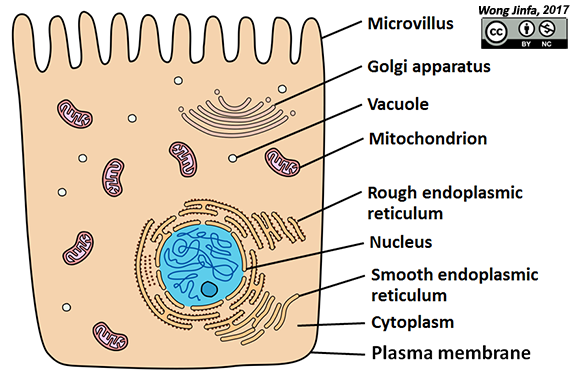
¶ State the characteristics and functions of the organelles and membrane structures found in the cell.
- Found within the cytoplasm
- A specialised subunit within the cell with a specific function
- Usually membrane bound
- Most require an electron microscope to be seen
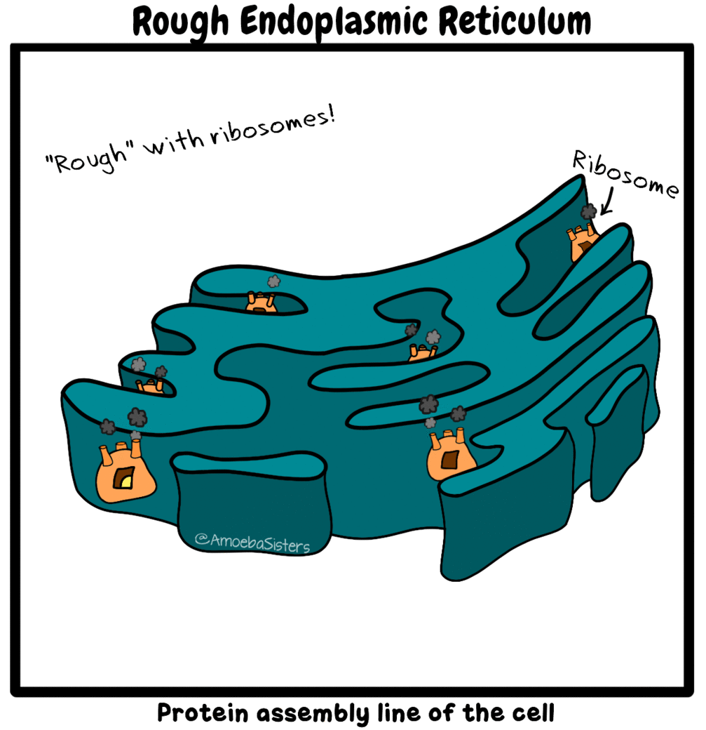
- Network of membrane-bound sacs continuous with nuclear envelope
- Rough Endoplasmic Reticulum
- Consist of flattened membrane-bound sacs called cisternae
- RER appears rough because small particles called ribosomes are attached to its outer surface
- Functions:
- Transport proteins made by ribosomes to the Golgi apparatus
- Modifies and folds proteins made by ribosomes into functional shapes
- Smooth Endoplasmic Reticulum
- Lacks ribosomes and has tubular sacs instead
- Functions:
- Synthesises substances such as fats (lipids) and steroids
- Converts harmful substances into harmless materials (detoxification)
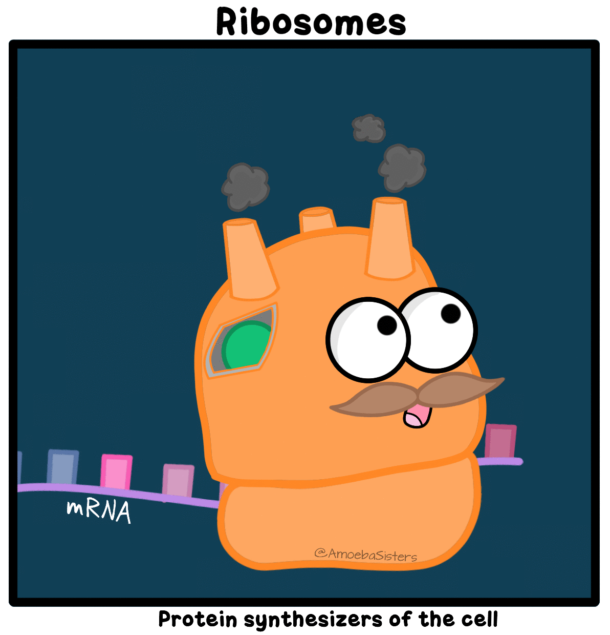
- Made of proteins and rRNA (ribosomal ribonucleic acids)
- May be attached to RER or lie freely in cytoplasm
- Function;
- Sites of protein synthesis
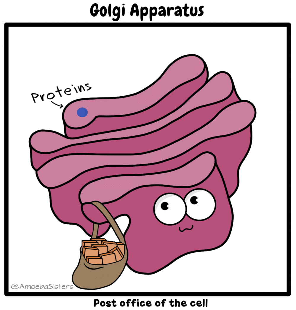
- Stacks of flattened / disc-shaped membrane-bound sacs
- Vesicles are seen fusing on one side of the Golgi membrane and pinching off from the opposite side
- Function:
- Modifies, stores and packages proteins in vesicles for secretion outside cell
- Secretory Pathway:
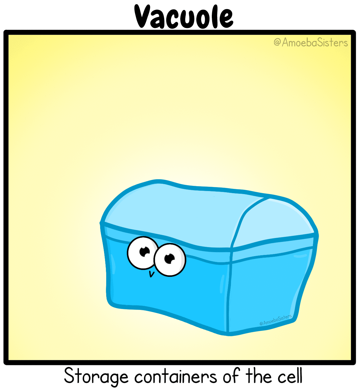
- Large vesicle which is filled with water that contains other dissolved substances
- The term vacuole, which means “empty,” refers to the fact that these organelles have no internal structure
- Animal Cells
- Have many small vacuoles for compartmentalisation
- Contain organic molecules or waste products for storage, transport, uptake or disposal
- Plant Cells
- One large central vacuole
- Contains cell sap: mostly water and dissolved salts, sugars, amino acids
- For storage or isolating harmful materials
- Give plant cells its structure by maintaining turgor pressure against cell wall
- May contain enzymes that are used for digestion
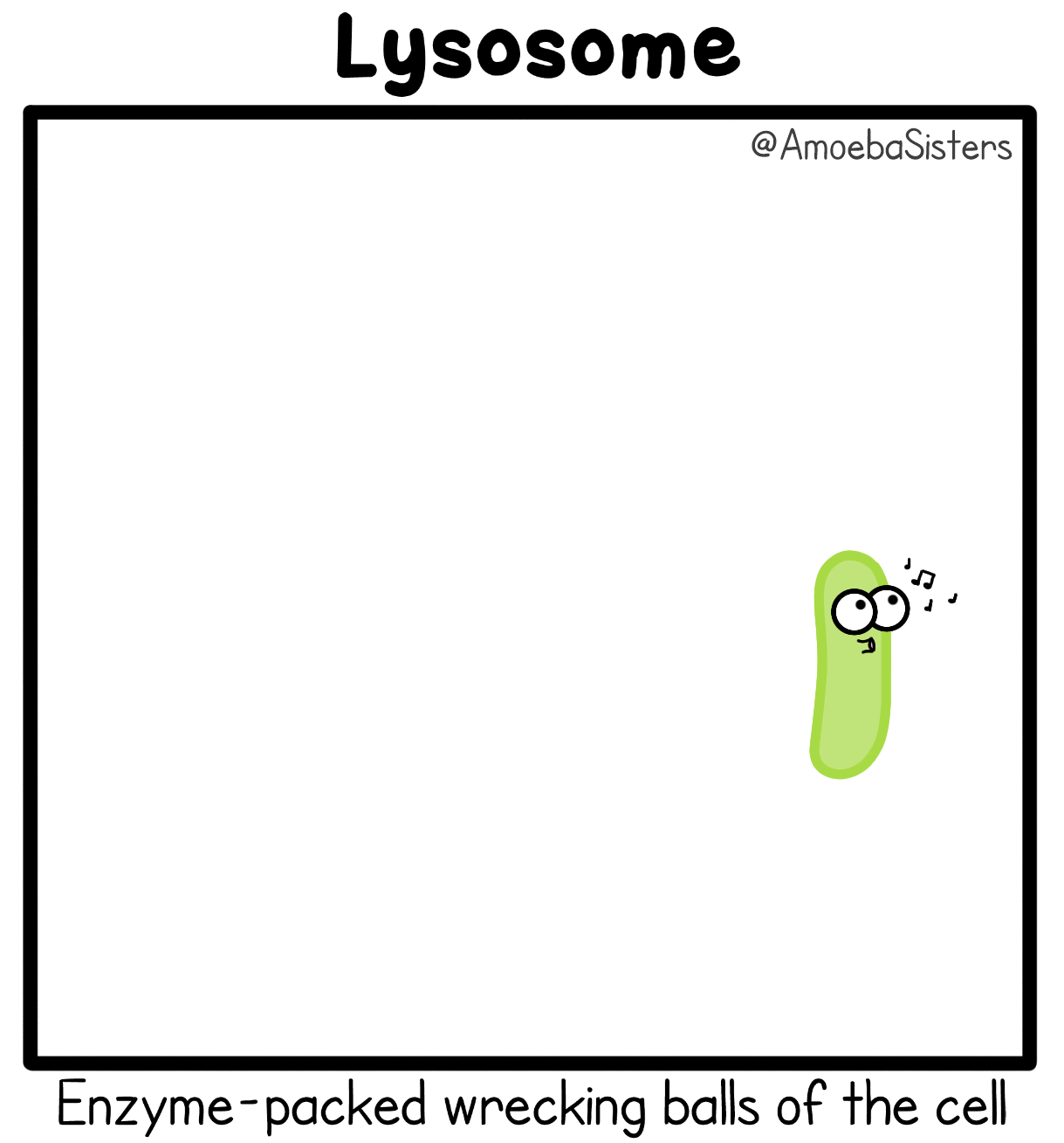
- In animal cells, they are small spherical vesicles
- In plant cells, large central vacuole may act as lysosomes
- Enzymes in the lysosomes are synthesized on RER & transported to golgi body where they form when they bud off
- Function:
- Contains digestive enzymes which breaks down proteins, nucleic acids & lipids
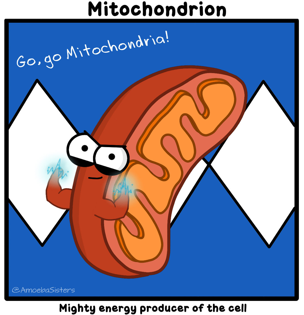
- Plural: Mitochondria
- Rod shaped organelle with a double membrane
- Highly in-folded inner membrane known as cristae increases surface area for ATP production
- Has its own DNA
- Function:
- Carries out cellular respiration, whereby glucose is broken down to release energy
- Energy is stored in the form of ATP (Adenosine Triphosphate), which is then transported to other parts of the cell that requires energy
- Disc-liked sac, visible with a light microscope
- Surrounded by two membranes which form the chloroplast envelope
- Inner membrane is continuous with stacks of flattened disc-sacs called thylakoids
- Has its own DNA
- Not all plant cells have chloroplasts
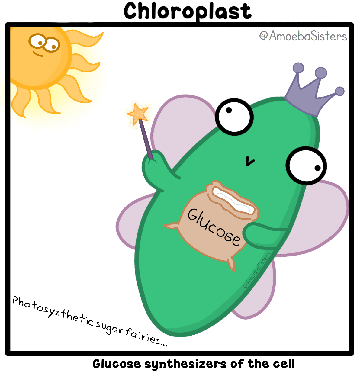
- Function:
- Carries out photosynthesis in plants by capturing light energy and storing them in glucose
- Click here for an animation on the theory
- Made up of microtubules, microfilaments and intermediate filaments
- Functions:
- Supports the cell and helps reinforce its shape
- Acts as tracks that guide motor proteins carrying organelles to their destination
- Found in animal cells, near nucleus, in a distinctly staining region of cytoplasm known as centrosomes
- Occur as a pair at right angles to each other
- One centriole consist of 9 triplets of microtubules in a single ring
- Organise spindle fibres which pulls chromosomes apart during cell division
- Give rise to basal bodies which anchor cilia & flagella
¶ Compare the structure of typical animal and plant cells.
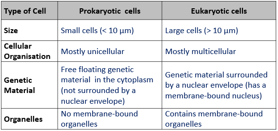
¶ State the main differences between prokaryotic and eukaryotic cells.
- Unicellular organisms contain all functions in a single cell to sustain life
- Multi-cellular organisms need to specialise so that they are able to perform the functions needed to sustain life more efficiently and effectively
- Multicellular organisms are made up of many different types of cells which perform different specific functions
- Differentiation is the process by which a cell becomes specialised for a specific function
- Benefits of cell specialisation
- Allows cells to perform their specific functions efficiently and more effectively
- Cells with different functions can combine to create more complex structures (tissues, organs) with new features or properties
- Allows many functions to be performed simultaneously
¶ Describe the relationship between cell function and cell structure of red blood cells in transport of oxygen, root hair cells in absorption, and epithelial cells of the small intestines in absorption.
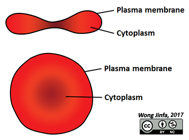
- Transports oxygen from lungs to all parts of the body
- Biconcave shape (thinner central portion)
- Optimises the flow of blood in blood vessels
- Increases surface area to volume ratio for more efficient gaseous exchange into and out of the cell
- Lack nucleus and most organelles
- More haemoglobin (iron-containing biomolecule that binds to oxygen) can be packed into the cytoplasm of the cell, enabling it to carry more oxygen
- Flexible and deformable membrane
- Allows it to squeeze through tiny capillaries.
- Are modified epidermal cells that aid in absorption of water at the roots
- Elongated shape
- Increases surface area to volume ratio for absorption of water and dissolved mineral salts at a faster rate
- Concentrated cell sap
- Gives cell a lower water potential than soil solution, and thus increase water uptake by osmosis
- Many mitochondria
- Releases energy for active transport of mineral salts
- The wall of the small intestines contains finger-like projections called villi
- Each villus is lined with a single layer of epithelial cells
- The epithelial cells of the small intestines have a highly infolded cell surface membrane on one side, which form protrusions called microvilli
- Microvilli
- Increases surface area to volume ratio for higher rate of absorption of digested food substances
- Many mitochondria
- Releases energy for active transport of digested food substance
- Protoplasm of cell is made up of three parts:
- Cell surface membrane
- Cytoplasm
- Nucleus